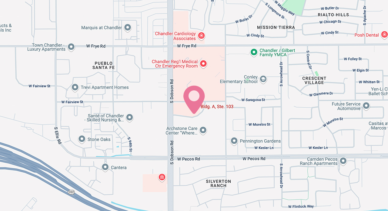
Address:
655 S Dobson Road, Building A, Unit 103
Chandler, AZ 85224


When Is Contrast-Enhanced Mammography Recommended?

Breast imaging has steadily evolved into a landscape of sharper detail, clearer contrasts, and more confident diagnoses. For patients in Scottsdale exploring contrast enhanced screening 3D and 2D mammograms, understanding when CEM is recommended can make the next step feel far less uncertain.
What Is Contrast-Enhanced Mammography?
CEM is performed in two main steps:
• A contrast agent - an iodine-based material - is injected into a vein.
• Mammography images are taken using both low-energy (similar to standard mammography) and high-energy exposures.
The combination creates images that show both the structure of breast tissue and the areas where contrast gathers. Because abnormal tissue can have increased blood supply, it may appear more prominently, helping radiologists detect issues earlier and with greater confidence.
Compared with MRI, CEM is faster, widely accessible, and typically more comfortable for patients, while still delivering high-quality diagnostic information.
When Standard Mammograms Show an Inconclusive Finding
A traditional 2D or 3D mammogram sometimes reveals a shadow, asymmetry, or area of distortion that requires clarification. Rather than immediately recommending a biopsy or MRI, clinicians may suggest CEM as a next step. The enhanced contrast can help:
• Confirm whether the area is suspicious
• Rule out false positives
• Reduce unnecessary procedures
This is particularly helpful during diagnostic follow-ups after a routine screening.
When You Have Dense Breast Tissue
Dense breast tissue - common among younger women and many over 40 - can hide abnormalities behind layers of overlapping tissue. CEM improves detectability by revealing contrast uptake, even in dense areas. Many patients prefer contrast enhanced screening 3D and 2D mammograms in Scottsdale because they feel more confident knowing their screening is equipped to “see through” density challenges.
When You Are at Higher Risk for Breast Cancer
Women who have a family history of breast cancer, certain genetic mutations, or other high-risk factors may benefit from enhanced imaging at regular intervals. CEM can be used in conjunction with annual mammography to strengthen early detection efforts, particularly in women who cannot undergo MRI.
When Treatment Planning Requires More Detail
CEM can reveal:
• Tumor size
• Tumor extent
• Multifocal or multicentric disease
• Whether a suspicious area has vascular activity suggesting malignancy
This information helps guide surgeons in planning lumpectomies or mastectomies and supports oncologists in evaluating how treatment is progressing.
Schedule Your Breast Imaging Today
Contrast-enhanced mammography is a remarkable tool for achieving clearer, more decisive breast imaging. For many women in Scottsdale, combining contrast enhanced screening 3D and 2D mammograms with additional diagnostic approaches - such as stereotactic biopsy when needed - creates a comprehensive roadmap to confident breast health decisions.
If you’re unsure whether contrast-enhanced mammography is right for you, our imaging specialists at URPrecious 3D Breast Ultrasound are here to help. Visit our office in Scottsdale, Arizona, or call (602) 878-750 to book a consultation today.

All Breast
Services

Keep
In Touch


© 2026 URPrecious Imaging. All rights Reserved - Accessibility Statement - Privacy Policy - Terms of Use - Sitemap
Powered by