
Address:
655 S Dobson Road, Building A, Unit 103
Chandler, AZ 85224


Mammograms
Advanced Breast Imaging
At URPrecious Imaging, we deliver exceptional breast imaging services that combine cutting-edge technology with compassionate, patient-centered care. Our goal is to make every mammogram a comfortable, confident experience while providing the highest standards of clinical excellence.
We provide AI at no additional patient cost
as we believe all patients need the equal and highest quality of care, when it comes to breast cancer screening and diagnosis.
Mammograms are interpreted using
DeepHealth SmartMammo™ AI,
Breast arterial calcification detection (Cardiovascular assessment) and
Tyrer-Cuzick lifetime breast cancer risk assessment
Same day results with a personalized radiologist consultation to discuss optimal early detection strategies tailored to your individual needs.
What Is a Mammogram?
A mammogram is a medical imaging test using low-dose X-rays to examine breast tissue. It helps detect early signs of breast cancer, often before any symptoms appear or can be felt. Most women begin regular screening mammograms at age 40, but the best schedule for testing depends on personal risk factors and medical history.
LOW DOSE
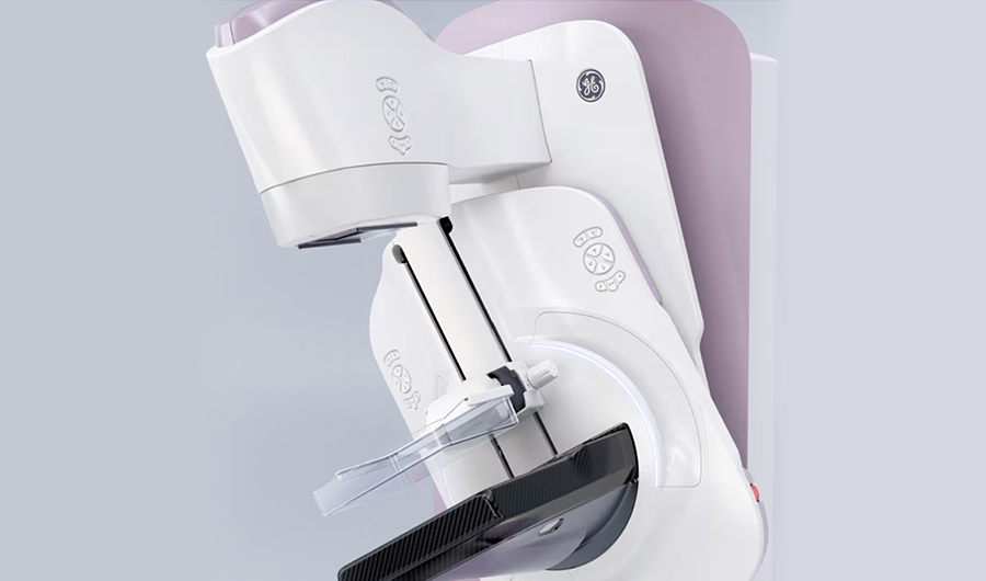
At URPrecious Imaging, the Senographe Pristina™ Mammography system delivers superior diagnostic accuracy with the lowest patient dose of all FDA approved Digital Breast Tomosynthesis systems.
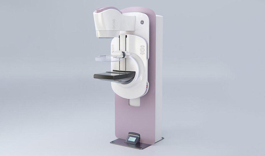
GE Pristina Via™
Experience mammography reimagined with GE HealthCare’s Senographe Pristina™, featuring low radiation dose and thoughtful design for superior image quality. Designed by women for women
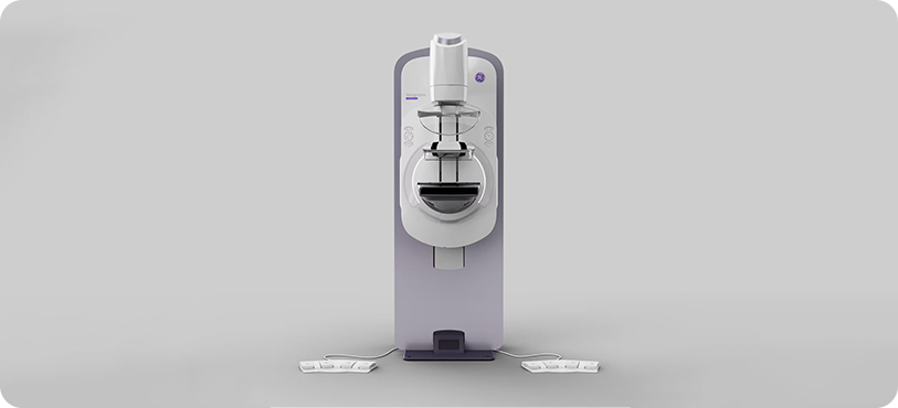
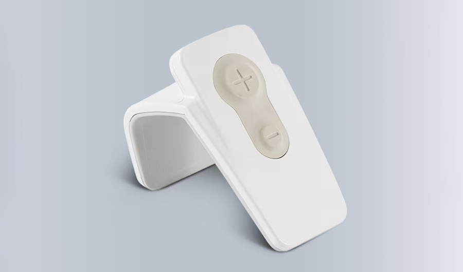
Under the radiographer’s supervision and guidance, our technology allows patients to control compression for a more comfortable mammographic experience.
Types of Mammograms
There are two primary types:
Screening mammogram: A routine test for women without any breast symptoms, intended to find cancer early.
Diagnostic mammogram: Performed if there are symptoms (like a lump, pain, or nipple discharge) or after an abnormal screening result. This exam takes more images for a closer look and may be combined with other tests.
Advances in Mammography
Digital mammography offers clearer computer images that can be adjusted for better viewing.
3D mammography (tomosynthesis) creates a more detailed, three-dimensional 1mm slices of images, especially helpful for dense breast tissue.
contrast-enhanced mammography (CEM) which provides vascular (blood flow) information, similar to breast MRI, but performed using iodinated contrast.
Advanced Breast Imaging
At URPrecious Imaging, we deliver exceptional breast imaging services that combine cutting-edge technology with compassionate, patient-centered care. Our goal is to make every mammogram a comfortable, confident experience while providing the highest standards of clinical excellence.
All mammograms at URPrecious Imaging are interpreted using DeepHealth SmartMammo™ AI, breast arterial calcification detection (Cardiovascular assessment) and Tyrer-Cuzick lifetime risk assessment. Each breast imaging appointment includes SAME DAY RESULTS with a personalized radiologist consultation to discuss optimal early detection strategies tailored to your individual needs.
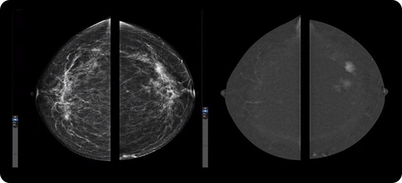
Contrast-Enhanced Mammography (CEM)
We offer Contrast-Enhanced Mammography (CEM) using iodinated contrast for advanced detection, demonstrating sensitivity rates of 96-97% and specificity of 84-87.5% in clinical studies—often matching or exceeding breast MRI performance while offering greater specificity in some cases. With accuracy up to 95% and superior sensitivity over standard mammography (e.g., 38% higher) and ultrasound, CEM is especially useful for patients with dense breasts, high-risk individuals, or those needing detailed lesion evaluation.
DeepHealth™ SmartMammo
We enhance clinical precision with AI driven detection by DeepHealth SmartMammo™ AI technology, which supports accurate breast density assessment and helps radiologists identify areas of interest with greater consistency. This advanced AI workflow streamlines interpretation and supports timely, personalized care—particularly valuable for those with dense breast tissue.
In the largest real-world analysis of AI-powered breast cancer screening in the U.S., a multistage AI-driven workflow from DeepHealth’s Breast Suite have shown to enable a 21% increase in breast cancer detection rate when compared to the standard of care . Additionally, Breast Suite’s Timely Alerts features uniquely integrates with Pristina Via to notify sites of potentially suspicious cases in minutes.
“Pristina Via and Breast Suite have transformed how we approach breast cancer detection,” said Dr. Meghna Krishnan, URPrecious Imaging, Arizona. “The cloud-based Viewer gives us instant access to images from anywhere, and the AI tools, like CAD for lesion detection and automated breast density assessment, can help us deliver fast and consistent results. It’s not just about speed, it’s about confidence. With Breast Suite, our team can make more accurate decisions without workflow disruptions, which ultimately means better care for our patients.”
The existing Breast Suite solution offered by GE HealthCare includes a cloud-first multi-modality Viewer, Cancer Detection, Automated Density Assessment, prioritized Worklist, Timely Alerts, and enhanced Reporting. The expanded collaboration will enable GE HealthCare to distribute new Breast Suite applications designed for compatibility with GE HealthCare’s mammography systems, including:
- ProFound Pro, which combines Cancer Detection, a clinical AI solution that offers automatic lesion localization and degree of suspicion that are effective in diverse populations and dense breast tissue, and Automated Density Assessment for consistent, automated density classification with a patient-centric, accurate density assessment of 2D or 3D mammograms to support objective diagnostic decisions.
- Safeguard Review: Optional AI-powered workflow that flags complex cases that may benefit from a secondary review. A large U.S. study demonstrated a 21% increase in cancer detection rate and a 23% increase in cancer detection rate for women with dense breasts with a multistage AI-driven workflow when compared to the standard of care.
Mammograms with Cardiovascular Interpretation
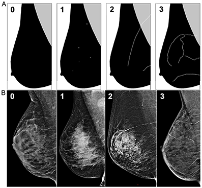
Our services extend beyond imaging with cardiovascular interpretation to detect and report breast arterial calcifications. Breast arterial calcification (BAC) on mammograms is a benign, often reported finding indicating medial arterial calcification, which correlates with cardiovascular disease (CVD) risk. It is commonly scored from Grade 0 (none) to Grade 3 (severe, widespread "tram-track" calcification) to assist in cardiovascular risk stratification, especially for women with high-risk cardiovascular factors.

Lifetime Risk Assessment with Genetic Testing
We also provide Tyrer-Cuzick risk assessment with radiologist consultation, plus hereditary cancer genetic testing through Myriad Genetics when clinically indicated, empowering you with a complete picture of your health.
What to Expect
During a mammogram, a technologist positions the breast on a platform and applies gentle compression. This spreads out the tissue for thinner, clearer X-ray images. The process may cause brief discomfort but is quick and safe.
Why Mammograms Matter
Mammograms can detect breast cancer years before physical symptoms develop, improving chances of successful treatment.
Routine mammograms are recommended every year for women ages from 40 and above, but higher-risk individuals may need to start earlier or get extra tests.
Early detection through mammography contributes to decreased morbidity and mortality related to breast cancer.
What Are Some Advantages Of Mammograms Over Ultrasound?
The physics of Ultrasound and mammogram is different from one another. Due to differences in breast composition, and differences in manifestation of different types of breast cancer such as calcifications, masses, architectural distortion, differences in blood flow or other forms, an appropriate usage of multiple modalities might be necessary to allow early detection of cancer. Mammogram is more sensitive than Ultrasound for detecting calcifications, architectural distortion and sometimes even masses based on their growth pattern. When breast cancer is detected early, then treatment may be minimal and prognosis the best. Routine annual mammographic screening has been shown to decrease breast cancer mortality by 40-50% since initiating mammographic screening in the USA.
We recommend following American College of Radiology appropriateness criteria and Society of breast imaging guidelines about what imaging may be appropriate for you based on your individual risk factors. Our fellowship trained breast imaging radiologist will be able to guide you about the recommended imaging for you, based on your risk factors.

All Breast
Services

Keep
In Touch


© 2026 URPrecious Imaging. All rights Reserved - Accessibility Statement - Privacy Policy - Terms of Use - Sitemap
Powered by